
OESOPHAGUS
INTRODUCTION
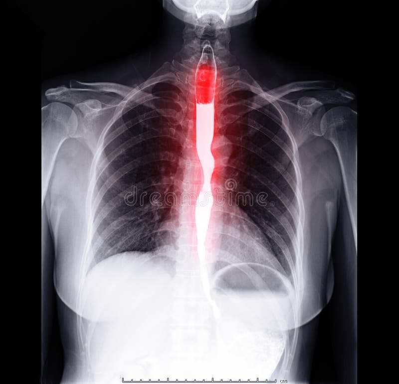
The oesophagus is about 25 cm long and about 2 cm in diameter and lies in the median plane in the thorax in front of the vertebral column behind the trachea and the heart. It is continuous with the pharynx above and just below the diaphragm it joins the stomach
It passes between muscle fibres of the diaphragm behind the central tendon at the level of the 10th thoracic vertebrae immediately the oesophagus has passed through the diaphragm it curves upward before opening into the stomach. This sharp angle is believed to be one of the factors that prevents the regurgitation gastric contents into the oesophagus.
The upper and lower ends of the oesophagus are closed by sphincters. The upper cricopharyngeal or upper oesophageal sphincter prevents air passing into the oesophagus during inspiration and the aspiration of oesophageal contents. The cardiac or lower oesophageal sphincter prevents the reflux of acid gastric contents into the oesophagus. There is no thickening of the circular muscle in this area and this sphincter is therefore 'physiological', I.e. this region can act as a sphincter without the presence of the anatomical features.
When intra-abdominal pressure is raised, e.g. during inspiration and defaecation, this tone of the lower oesophageal sphincter increases. There is an added pinching effect by the contracting muscle fibres of the diaphragm.
STRUCTURE OF THE OESOPHAGUS
There are four layers of tissues , as the oesophagus is almost entirely in the thorax the outer covering, the adventitia, consists of elastic fibrous tissue that attaches the oesophagus to the surrounding structures. The proximal third is lined by stratified squamous epithelium and the distal third by columnar epithelium. The middle third is lined by a mixture of the two.
FUNCTIONS OF THE OESOPHAGUS
Formation of bolus
When food is taken into the mouth it is chewed by the teeth and moved around the mouth by the tongue and muscles of the cheeks. It is mixed with saliva and formed into a soft mass or bolus ready for swallowing. The length of time that food remains in the mouth largely depends on the consistency of the food. Some food need to be chewed longer than others before the individual feels that the bolus is ready for swallowing.
Swallowing
There are three stages-
Oral stage
With the mouth closed, the voluntary muscles of the tongue and cheeks push the bolus backwards into the pharynx.
Pharyngeal stage
2. the muscle of the pharynx are stimulated by a reflex action initiated in the walls of the oropharynx and coordinated by the swallowing centre in the medulla. Involuntary contraction of these muscle propels the bolus down into the oesophagus. All other routes that the bolus could take are closed. The soft palate rises up and closes off the nasopharynx; the tongue and the pharyngeal folds block the way back into the mouth; and the larynx is lifted up and forward so that its opening is occluded by the overhanging epiglottis preventing entry into the airway(trachea)
oesophageal stage.
The presence of the bolus in the pharynx stimulates a wave of peristalsis that peoples the bolus through the oesophagus to the stomach. Peristaltic waves pass along the oesophagus only after swallowing begins. Otherwise the walls are relaxed.
Other factors preventing gastric reflux include:
BLOOD SUPPLY
Arterial
The thoracic region is supplied mainly by the paired oesophageal arteries, branches from the thoracic aorta. The abdominal region is supplied by branches from the inferior phrenic arteries and the left gastric branch of the coeliac artery.
Venous drainage
Venous drainage From the thoracic region venous drainage is into the azygos and hemiazygos veins. The abdominal part drains into the left gastric vein . There is a venous plexus at the distal end that links the upward and downward venous drainage, I.e. the general and portal circulation


0 Comments